Understanding the Spine, Spinal Stenosis, and Spinal Nerve Compression
Ryan Klopfer • March 26, 2024
Table of Contents
- Understanding the anatomy of the spine
- The lumbar vertebrae
- Muscles associated with the lumbar spine
- Ligaments of the lumbar spine
- Congenital anomalies of the lumbar spine
- Spinal stenosis or spinal nerve compression
- Causes of spinal stenosis
- Symptoms and effects of spinal stenosis
- Is spinal decompression right for you?
- Share This Post
- Georgia Upper Cervical Chiropractic
Did You Know That Your Anatomy and Environmental Factors Affect Your Spine?

Understanding the anatomy of the spine
The spine has the following key components:
The lumbar vertebrae
The lumbar spine consists of five vertebrae, labeled L1 through L5, which are the largest and strongest in the vertebral column. They have a characteristic kidney-shaped body, thicker and more robust than those in the thoracic and cervical regions, designed to support more weight.
- Intervertebral Discs: Each lumbar vertebra is separated by an intervertebral disc, composed of a tough, fibrous outer layer called the annulus fibrosus and a soft, gel-like center called the nucleus pulposus. These discs act as shock absorbers and provide flexibility to the spine.
- Spinous and Transverse Processes: Each vertebra has a spinous process projecting posteriorly and two transverse processes projecting laterally. These provide attachment points for muscles and ligaments.
- Articular Processes: Superior and inferior articular processes form facet joints with adjacent vertebrae, facilitating controlled movement and providing stability.
- Intervertebral Foramina: The intervertebral foramina, also known as neural foramina, are openings located between adjacent vertebrae. They are formed by the notches on the pedicles of the vertebrae above and below. In the lumbar spine, there are pairs of these foramina between each set of vertebrae.
i. Function and Significance: These foramina serve as passageways for nerves, blood vessels, and lymphatics. They play a crucial role in the nervous system, as they are the points through which the spinal nerves and blood vessels exit the spinal canal and extend to other parts of the body. These nerve roots branch from the spinal cord and are named for the vertebra above (e.g., the nerve root passing between the L4 and L5 vertebrae is named L4).
ii. Contents of the Foramina: Spinal Nerves: These nerves are responsible for motor, sensory, and autonomic functions of the body parts they innervate, including the lower extremities, parts of the abdomen, and some regions of the back.
iii. Blood Vessels: The foramina also contain important blood vessels that supply the spinal cord, vertebrae, and other spinal structures. These include segmental arteries, which branch off from larger systemic arteries, and accompanying veins.
iv. Lymphatics and Adipose Tissue: Along with nerves and blood vessels, the foramina also contain lymphatic vessels and a small amount of adipose tissue. The adipose tissue acts as a protective cushion for the nerve roots.
Clinical Significance: The size and shape of the lumbar intervertebral foramina can change due to various pathological conditions, such as disc herniation, spinal stenosis, or spondylolisthesis. These changes can lead to nerve root compression, causing symptoms like pain, numbness, tingling or weakness in the areas innervated by the affected nerves (often referred to as radiculopathy).
Diagnostic imaging, such as MRI or CT scans, is often used to evaluate the foramina and diagnose conditions that affect them. In summary, the intervertebral foramina in the lumbar spine are critical structures that facilitate the exit of spinal nerves and blood vessels from the spinal column. Their integrity is vital for the proper functioning of the lower part of the body, and any pathology affecting these foramina can have significant clinical implications.
Muscles associated with the lumbar spine
- Erector Spinae: This group of muscles runs along the back from the sacrum to the cervical spine and is critical for maintaining posture and erect position. It consists of three columns of muscles: iliocostalis, longissimus, and spinalis.
- Quadratus Lumborum: Located on either side of the lumbar spine, it aids in lateral flexion of the vertebral column and stabilizes the pelvis.
- Psoas Major: Originating from the lumbar vertebrae and inserting into the femur, this muscle flexes the hip joint and stabilizes the lumbar region.
- Multifidus: This deep muscle runs along the vertebral column and provides stabilization and support to the spine during movement.
- Latissimus Dorsi: Although primarily a thoracic muscle, it attaches to the lumbar region and assists in movements of the trunk.
Ligaments of the lumbar spine
Each of these structures plays a specific and crucial role in the overall function of the lumbar spine, ensuring stability, flexibility, and protection for the spinal cord and nerves while supporting the body's weight. Injuries, strain, or degenerative changes in any of these components can lead to back pain and other lumbar spine-related issue.
- Anterior Longitudinal Ligament: This runs along the anterior surface of the vertebral bodies from the cervical to the lumbar region, limiting extension.
- Posterior Longitudinal Ligament: Running within the vertebral canal along the posterior side of the vertebral bodies, it restricts flexion.
- Ligamentum Flavum: Connecting the laminae of adjacent vertebrae, it preserves the spinal canal's shape and assists in straightening the back after flexing.
- Interspinous Ligaments: These are situated between the spinous processes of the vertebrae, aiding in the stabilization of the spine.
- Supraspinous Ligament: This strong ligament connects the tips of the spinous processes from the seventh cervical vertebra to the sacrum, limiting flexion.
- Iliolumbar Ligaments: These extend from the transverse processes of the lumbar vertebrae to the iliac crests, providing stability to the lumbo-sacral junction.
Congenital anomalies of the lumbar spine
Anomanies are variations from the normal anatomy that occur during the development of the spine in the womb. These anomalies can range from mild to severe and can impact the structure and function of the spine.
Some of the most common congenital anomalies in the lumbar spine include:
- Spina Bifida: Spina bifida is a neural tube defect characterized by the incomplete closing of the backbone and membranes around the spinal cord. There are several types, ranging from mild (spina bifida occulta, where there is a small gap in one or more of the vertebrae but no opening or sac on the back) to severe (such as myelomeningocele, where the spinal canal remains open along several vertebrae in the lower or middle back, allowing the spinal cord and membranes to protrude through an opening).
- Spondylolisthesis: This condition occurs when a vertebra slips forward over the one below it. Congenital spondylolisthesis typically results from a malformation of the lumbosacral joint, the joint between the last lumbar vertebra and the first sacral segment.
- Hemivertebra: A hemivertebra is a wedge-shaped vertebra that can cause an abnormal angle or curve in the spine (scoliosis). It occurs when one side of a vertebra fails to form normally, leading to a unilateral growth and asymmetrical spinal column.
- Congenital Scoliosis: Unlike idiopathic scoliosis that develops later in life, congenital scoliosis is present at birth. It is caused by a malformation of the spine during development, such as the failure of vertebrae to form completely or the formation of extra vertebrae.
- Sacral Agenesis: This rare, severe condition involves the underdevelopment or absence of part or all of the sacrum (the lower spine) and occasionally the lumbar spine. It is often associated with abnormalities of lower limbs and lower parts of the nervous system.
- Transitional Vertebrae: A transitional vertebra occurs when a vertebra at the lower spine (usually at the lumbosacral junction) has characteristics of both a lumbar and a sacral vertebra. The most common type is a lumbarized sacrum where the first sacral vertebra forms as a lumbar vertebra.
These congenital anomalies can vary greatly in terms of symptoms and severity. Some individuals may remain asymptomatic and unaware of their condition, while others may experience pain, neurological symptoms, or significant deformity. Diagnosis is typically made through imaging studies such as X-rays, CT scans, or MRI. Treatment depends on the specific anomaly and its effects and may range from observation and physical therapy to surgical intervention in more severe cases.
Spinal stenosis or spinal nerve compression
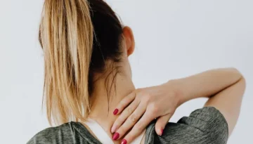
Spinal stenosis is a condition characterized by the narrowing of spaces within your spine, which can put pressure on the nerves that travel through the spine. This condition most commonly affects the lower back and the neck.
There are two main types of spinal stenosis:
- Cervical Stenosis: Involves the narrowing of the spine in the neck.
2.Lumbar Stenosis: Involves the narrowing of the spine in the lower back, which is more common.
Causes of spinal stenosis
The causes of spinal stenosis, also known as nerve compression, can include:
- Aging: With age, the body's ligaments can thicken, and bones and joints can enlarge, leading to a narrowing of the spinal canal.
- Osteoarthritis: The wear and tear damage associated with osteoarthritis can prompt the formation of bone spurs, which can grow into the spinal canal.
- Herniated Discs: The soft cushions between vertebrae may dry out and crack, allowing some of the soft inner material to escape and press on the spinal cord or nerves
- Injuries: Accidents and trauma can cause fractures or dislocations of one or more vertebrae, which can damage the contents of the spinal canal.
- Tumors: Abnormal growths can form inside the spinal cord, within the membranes that cover the spinal cord, or in the space between the spinal cord and vertebrae.
Symptoms and effects of spinal stenosis
The symptoms of spinal stenosis and nerve compression can vary depending on the location and severity of the stenosis. Common symptoms include:
- Pain in the neck or back: Depending on where the stenosis is located.
- Numbness, tingling, or weakness: These sensations can occur in the arms, hands, legs, or feet.
- Problems with walking and balance: Lumbar stenosis can cause cramping or pain in the legs, especially when walking or standing for long periods.
In severe cases, bladder or bowel dysfunction: This is a rare but serious symptom and requires immediate medical attention. Now that you know about the signs and symptoms, learn about the treatment options.
Is spinal decompression right for you?
Spinal decompression is an effective treatment for relieving pressure on the spinal cord or nerves. There are two types: surgical and non-surgical. Surgical decompression involves procedures to remove parts of bone or disc material pressing on nerves, while non-surgical decompression uses motorized traction to gently stretch the spine, reducing pressure and promoting healing.
Ready to book an appointment with Georgia Upper Cervical?
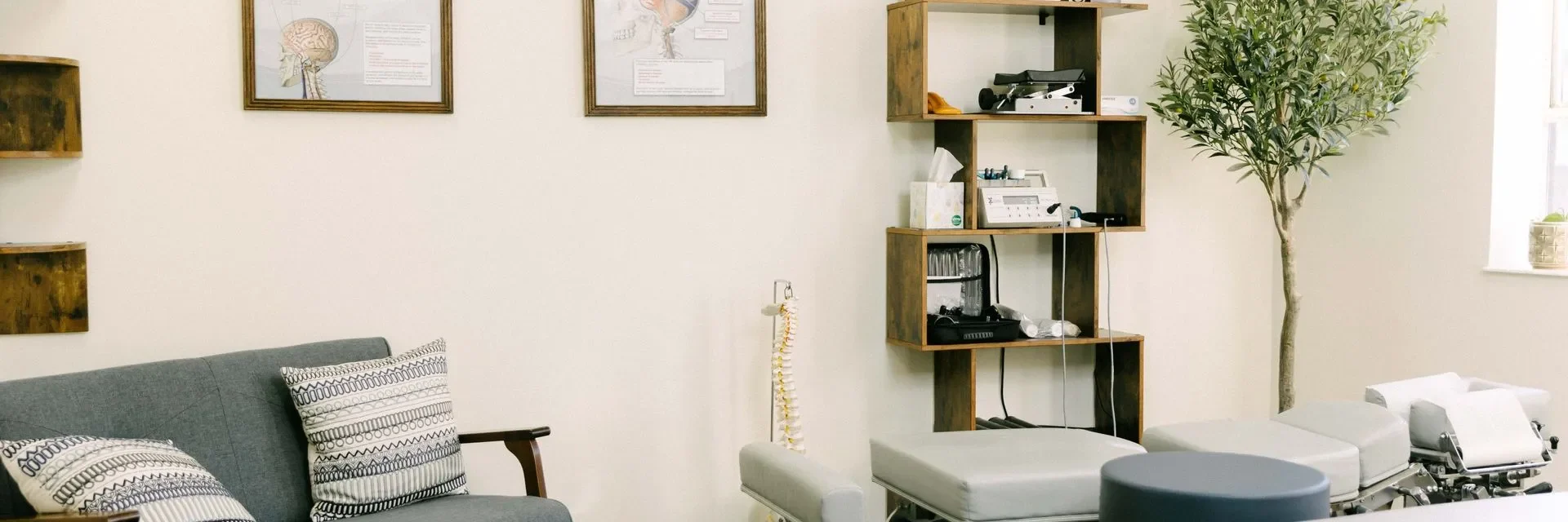
If you suffer from spinal stenosis or spinal compression, Georgia Upper Cervical is here to provide comprehensive assistance. Located in Ball Ground, GA, our dedicated team is committed to empowering you with knowledge and understanding of these conditions, enabling you to take proactive steps towards a better, stronger, and happier life. With a focus on patient-centered care, we prioritize your well-being and strive to equip you with the tools and support necessary to navigate through the challenges posed by spinal stenosis and compression.
At Georgia Upper Cervical, we believe that every individual deserves the opportunity to live life to the fullest, free from the constraints of spinal discomfort. Our expert team utilizes advanced techniques and state-of-the-art equipment to deliver tailored solutions designed to address your unique needs effectively. If you're ready to take control of your spinal health and embark on a path towards lasting relief and vitality, schedule an appointment with Georgia Upper Cervical today. Let us partner with you on your journey to optimal well-being.